New hope for rehabilitation and recovery for patients with sight loss
Researchers have used MRI imaging to map visual brain activity in stroke survivors with sight loss that gives new hope for rehabilitation and recovery.
Scientists from the University of Nottingham have revealed new insights by combining data from clinical sight tests with brain imaging to precisely map the areas of the brain affected by sight loss. This allows identification of visual brain areas where function could potentially be improved with rehabilitation. The research, which was funded by the charity Fight for Sight has been published today in Frontiers in Neuroscience.
Every year around 150,000 people in the UK have a stroke with roughly 30% experiencing some kind of sight loss as a result. Visual field loss is a common and devastating complication of cerebral strokes. This type of sight loss, called hemianopia, affects one side of a person's vision and is caused by damage to the visual pathway in the brain.
The challenge for patients with visual field loss is their active participation in circulation, and some might never be able to drive their car anymore safely. Most countries test stroke patients on their ability to drive.
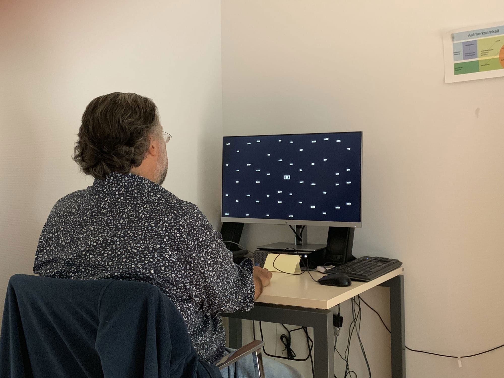
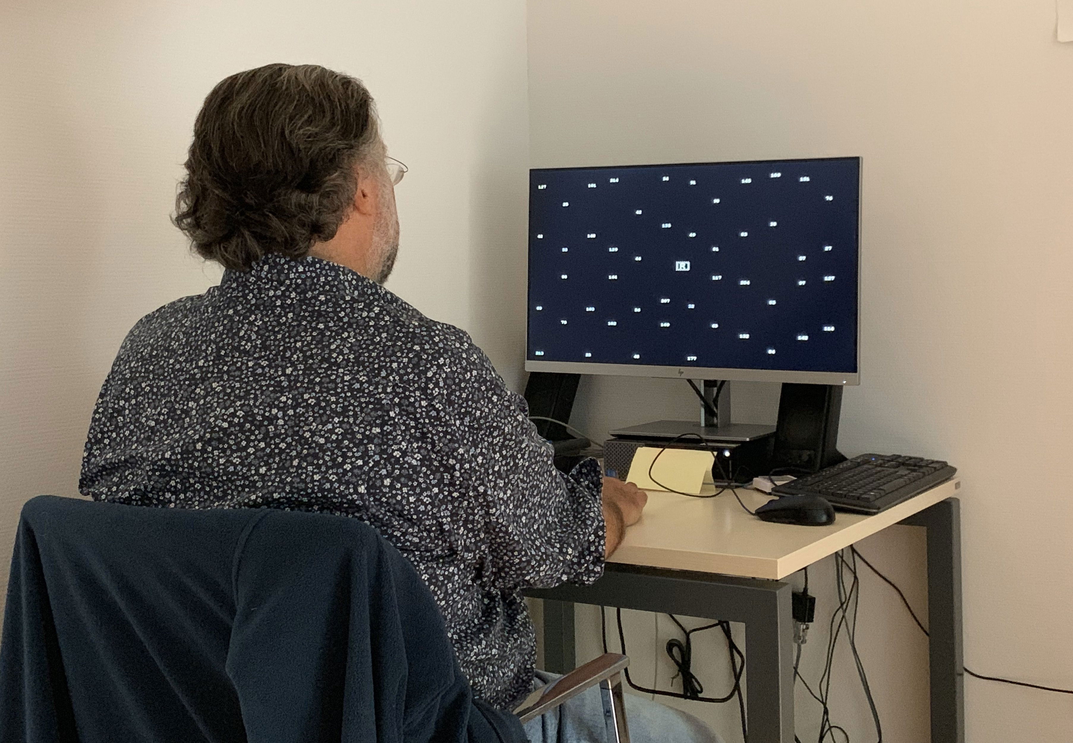
A visual field test called perimetry is the current gold standard for measuring residual visual field coverage. The picture is showing Mr. Steven Saltzman, President of the Monaco AVC Association performing a residual visual test in the Rehabilitation Clinic in Kilchberg, Switzerland. www.zurzachcare.ch
Read the full article:
https://www.sciencedaily.com/releases/2022/01/220106105957.htm


